Review


human bladder epithelial cell line 5637  (ATCC)


Bioz Verified Symbol ATCC is a verified supplier
Bioz Manufacturer Symbol ATCC manufactures this product  
  • Logo
  • About
  • News
  • Press Release
  • Team
  • Advisors
  • Partners
  • Contact
  • Bioz Stars
  • Bioz vStars
  • 94

    Structured Review

    ATCC human bladder epithelial cell line 5637
    Human Bladder Epithelial Cell Line 5637, supplied by ATCC, used in various techniques. Bioz Stars score: 94/100, based on 80 PubMed citations. ZERO BIAS - scores, article reviews, protocol conditions and more
    https://www.bioz.com/product/5637+human+bladder+epithelial+carcinoma+cell+line/ME-180%3A+Cervical+Carcinoma%3B+Human/pmc11724981__41467_2025_55982_MOESM4_ESM-53-12-18
    Average 94 stars, based on 80 article reviews
    human bladder epithelial cell line 5637 - by Bioz Stars, 2026-09
    94/100 stars

    Images

    Related Articles

    Cell Culture:

    Article Title: Dynamic persistence of UPEC intracellular bacterial communities in a human bladder-chip model of urinary tract infection
    Article Snippet: .. Cell culture of human bladder epithelial and bladder endothelial cells The 5637 human bladder epithelial carcinoma cell line (ATCC, HTB-9) was cultured in RPMI 1640 medium supplemented with 10% Fetal Bovine Serum (FBS) as recommended by the supplier. ..

    Article Title: Dynamic persistence of UPEC intracellular bacterial communities in a human bladder-chip model of urinary tract infection
    Article Snippet: .. The 5637 human bladder epithelial carcinoma cell line (ATCC, HTB-9) was cultured in RPMI 1640 medium supplemented with 10% Fetal Bovine Serum (FBS) as recommended by the supplier. ..

    Infection:

    Article Title: Advances in experimental bladder models: bridging the gap between in vitro and in vivo approaches for investigating urinary tract infections
    Article Snippet: Mouse bladder organoids from C57BL/6 wild-type or mT/mG mice , UPEC , To investigate how early invasion of the bladder wall by solitary bacteria can protect UPEC from the effects of antibiotics and the response of neutrophil swarms, using an organoid model , In bladder organoid model, solitary bacteria invaded deeper layers of the bladder wall early in infection, independent of the formation and rupture of intracellular bacterial communities (IBCs). These solitary bacteria, which resemble quiescent intracellular reservoirs (QIRs), evade destruction by antibiotics and neutrophils. They are distinct in morphology from bacteria within IBCs, suggesting that QIR-like bacteria can form early during infection. , [ ] . .. • 5637 human bladder epithelial carcinoma cell line (procured from ATCC HTB-9TM) • Human primary bladder epithelial cells • Human Bladder Micro-vascular Endothelial cells (HMVEC-Bd) , UPEC strain CFT073 , To develop and characterize a bladder-chip model that mimics bladder structure by co-culturing human bladder epithelial cells with bladder microvascular endothelial cells, using a device that exposes epithelial cells to urine and endothelial cells to culture media , Neutrophils rapidly recruited from the vascular channel to infection sites formed swarms and extracellular traps, but this did not prevent the formation of intracellular bacterial communities (IBCs). Antibiotics showed delayed effectiveness in eliminating bacteria within IBCs, and in some cases, bacteria were not eradicated at all. During recovery periods, bacteria rapidly proliferated in IBCs, leading to new infection sites through bacterial shedding and host cell exfoliation. , [ ] . .. HBLAK human bladder progenitor cells , UPEC strains UTI89, CFT073, E. coli 83,972 Enterococcus faecalis EF36, EF77 and Streptococcus agalactiae , To develop and utilize an immune-responsive three-dimensional urine-tolerant human urothelial model to study UTIs , 3D urine-tolerant human urothelial model (3D-UHU) was stratified into 7–8 layers with distinct cell types, after 18–20 days. The apical surface differentiated into CD227 + umbrella-like cells expressing key urothelial markers (uroplakin-1 A, II, III, and cytokeratin 20) and a glycosaminoglycan layer, while intermediate and basal cells (CD271+) were present underneath. The model showed effective barrier function and expressed proteins like E-cadherin, claudin-1 and − 3, and ZO-1. Infection with both Gram-negative and Gram-positive bacteria led to increased pro-inflammatory cytokines and chemokines, mimicking human UTI. This model offered potential for exploring host-pathogen interactions and host urothelial immune responses. , [ ] .

    Bacteria:

    Article Title: Advances in experimental bladder models: bridging the gap between in vitro and in vivo approaches for investigating urinary tract infections
    Article Snippet: Mouse bladder organoids from C57BL/6 wild-type or mT/mG mice , UPEC , To investigate how early invasion of the bladder wall by solitary bacteria can protect UPEC from the effects of antibiotics and the response of neutrophil swarms, using an organoid model , In bladder organoid model, solitary bacteria invaded deeper layers of the bladder wall early in infection, independent of the formation and rupture of intracellular bacterial communities (IBCs). These solitary bacteria, which resemble quiescent intracellular reservoirs (QIRs), evade destruction by antibiotics and neutrophils. They are distinct in morphology from bacteria within IBCs, suggesting that QIR-like bacteria can form early during infection. , [ ] . .. • 5637 human bladder epithelial carcinoma cell line (procured from ATCC HTB-9TM) • Human primary bladder epithelial cells • Human Bladder Micro-vascular Endothelial cells (HMVEC-Bd) , UPEC strain CFT073 , To develop and characterize a bladder-chip model that mimics bladder structure by co-culturing human bladder epithelial cells with bladder microvascular endothelial cells, using a device that exposes epithelial cells to urine and endothelial cells to culture media , Neutrophils rapidly recruited from the vascular channel to infection sites formed swarms and extracellular traps, but this did not prevent the formation of intracellular bacterial communities (IBCs). Antibiotics showed delayed effectiveness in eliminating bacteria within IBCs, and in some cases, bacteria were not eradicated at all. During recovery periods, bacteria rapidly proliferated in IBCs, leading to new infection sites through bacterial shedding and host cell exfoliation. , [ ] . .. HBLAK human bladder progenitor cells , UPEC strains UTI89, CFT073, E. coli 83,972 Enterococcus faecalis EF36, EF77 and Streptococcus agalactiae , To develop and utilize an immune-responsive three-dimensional urine-tolerant human urothelial model to study UTIs , 3D urine-tolerant human urothelial model (3D-UHU) was stratified into 7–8 layers with distinct cell types, after 18–20 days. The apical surface differentiated into CD227 + umbrella-like cells expressing key urothelial markers (uroplakin-1 A, II, III, and cytokeratin 20) and a glycosaminoglycan layer, while intermediate and basal cells (CD271+) were present underneath. The model showed effective barrier function and expressed proteins like E-cadherin, claudin-1 and − 3, and ZO-1. Infection with both Gram-negative and Gram-positive bacteria led to increased pro-inflammatory cytokines and chemokines, mimicking human UTI. This model offered potential for exploring host-pathogen interactions and host urothelial immune responses. , [ ] .

    other:

    Article Title: Dynamic persistence of UPEC intracellular bacterial communities in a human bladder-chip model of urinary tract infection
    Article Snippet: 5637 human bladder epithelial carcinoma cell line (procured from ATCC, HTB-9TM).


    Similar Products

    94
    ATCC human bladder epithelial cell line 5637
    Human Bladder Epithelial Cell Line 5637, supplied by ATCC, used in various techniques. Bioz Stars score: 94/100, based on 1 PubMed citations. ZERO BIAS - scores, article reviews, protocol conditions and more
    https://www.bioz.com/product/5637+human+bladder+epithelial+carcinoma+cell+line/ME-180%3A+Cervical+Carcinoma%3B+Human/pmc11724981__41467_2025_55982_MOESM4_ESM-53-12-18
    Average 94 stars, based on 1 article reviews
    human bladder epithelial cell line 5637 - by Bioz Stars, 2026-09
    94/100 stars
      Buy from Supplier

    97
    ATCC 5637 human bladder epithelial carcinoma cell line
    Comparison of different organoids used as bladder models
    5637 Human Bladder Epithelial Carcinoma Cell Line, supplied by ATCC, used in various techniques. Bioz Stars score: 97/100, based on 1 PubMed citations. ZERO BIAS - scores, article reviews, protocol conditions and more
    https://www.bioz.com/product/5637+human+bladder+epithelial+carcinoma+cell+line/5637/pmc11418205-30-0-10
    Average 97 stars, based on 1 article reviews
    5637 human bladder epithelial carcinoma cell line - by Bioz Stars, 2026-09
    97/100 stars
      Buy from Supplier

    94
    ATCC bladder epithelial cell line 5637
    Comparison of different organoids used as bladder models
    Bladder Epithelial Cell Line 5637, supplied by ATCC, used in various techniques. Bioz Stars score: 94/100, based on 1 PubMed citations. ZERO BIAS - scores, article reviews, protocol conditions and more
    https://www.bioz.com/product/5637+human+bladder+epithelial+carcinoma+cell+line/ME-180%3A+Cervical+Carcinoma%3B+Human/pmc10245908-81-2-7
    Average 94 stars, based on 1 article reviews
    bladder epithelial cell line 5637 - by Bioz Stars, 2026-09
    94/100 stars
      Buy from Supplier

    97
    ATCC htb9 human bladder epithelial carcinoma cell line
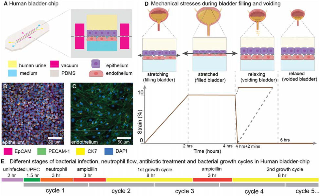
    (A) Schematic of the human bladder-chip with co-culture of the <t>HTB9</t> human bladder <t>epithelial</t> cell line (epithelium, top) and primary human bladder microvascular endothelial cells (endothelial, bottom) on either side of the stretchable and porous membrane. Pooled human urine diluted in PBS and endothelial cell medium were perfused in the apical and vascular channels respectively to mimic bladder physiology. A negative pressure in the ‘vacuum’ channels (magenta) on either side of the main channel was applied to stretch the porous membrane to mimic stretching of the bladder. ( B , C ) Immunofluorescence staining of confluent epithelial and endothelial cell monolayers (anti-EpCAM (magenta) and anti-CK7 (yellow) for the epithelial cells and anti-PECAM-1 (green) for the endothelial cells) in an uninfected control chip. Some endothelial cells also stained positive for CK7. Cell nuclei were labeled with DAPI (azure). ( D ) Schematic of the reconstitution of the bladder filling and voiding cycle via stretching of the membrane with a duty cycle of 6 hours. The cycle consisted of a linear increase in strain through stretching of the membrane ( filling bladder , 0 to 2 hours), maintenance of the membrane under stretch ( filled bladder , 2 to 4 hours), a quick relaxation of applied strain over 2 minutes ( voiding bladder , 4:02 hours) and maintenance without applied strain ( voided bladder , 4:02 hours to 6hours). ( E ) An overview of the timeline of the experimental protocol including infection, addition of neutrophils via the vascular channel, and two cycles of antibiotic treatment interspersed by two bacterial growth cycles. The consecutive bladder duty cycles are indicated.
    Htb9 Human Bladder Epithelial Carcinoma Cell Line, supplied by ATCC, used in various techniques. Bioz Stars score: 97/100, based on 1 PubMed citations. ZERO BIAS - scores, article reviews, protocol conditions and more
    https://www.bioz.com/product/5637+human+bladder+epithelial+carcinoma+cell+line/5637/bio_rxiv__2021__01__03__424836-221-1-10
    Average 97 stars, based on 1 article reviews
    htb9 human bladder epithelial carcinoma cell line - by Bioz Stars, 2026-09
    97/100 stars
      Buy from Supplier

    Image Search Results


    Comparison of different organoids used as bladder models

    Journal: BMC Urology

    Article Title: Advances in experimental bladder models: bridging the gap between in vitro and in vivo approaches for investigating urinary tract infections

    doi: 10.1186/s12894-024-01590-w

    Figure Lengend Snippet: Comparison of different organoids used as bladder models

    Article Snippet: • 5637 human bladder epithelial carcinoma cell line (procured from ATCC HTB-9TM) • Human primary bladder epithelial cells • Human Bladder Micro-vascular Endothelial cells (HMVEC-Bd) , UPEC strain CFT073 , To develop and characterize a bladder-chip model that mimics bladder structure by co-culturing human bladder epithelial cells with bladder microvascular endothelial cells, using a device that exposes epithelial cells to urine and endothelial cells to culture media , Neutrophils rapidly recruited from the vascular channel to infection sites formed swarms and extracellular traps, but this did not prevent the formation of intracellular bacterial communities (IBCs). Antibiotics showed delayed effectiveness in eliminating bacteria within IBCs, and in some cases, bacteria were not eradicated at all. During recovery periods, bacteria rapidly proliferated in IBCs, leading to new infection sites through bacterial shedding and host cell exfoliation. , [ ] .

    Techniques: Comparison, Mutagenesis, Cell Culture, Expressing, Bacteria, Infection

    (A) Schematic of the human bladder-chip with co-culture of the HTB9 human bladder epithelial cell line (epithelium, top) and primary human bladder microvascular endothelial cells (endothelial, bottom) on either side of the stretchable and porous membrane. Pooled human urine diluted in PBS and endothelial cell medium were perfused in the apical and vascular channels respectively to mimic bladder physiology. A negative pressure in the ‘vacuum’ channels (magenta) on either side of the main channel was applied to stretch the porous membrane to mimic stretching of the bladder. ( B , C ) Immunofluorescence staining of confluent epithelial and endothelial cell monolayers (anti-EpCAM (magenta) and anti-CK7 (yellow) for the epithelial cells and anti-PECAM-1 (green) for the endothelial cells) in an uninfected control chip. Some endothelial cells also stained positive for CK7. Cell nuclei were labeled with DAPI (azure). ( D ) Schematic of the reconstitution of the bladder filling and voiding cycle via stretching of the membrane with a duty cycle of 6 hours. The cycle consisted of a linear increase in strain through stretching of the membrane ( filling bladder , 0 to 2 hours), maintenance of the membrane under stretch ( filled bladder , 2 to 4 hours), a quick relaxation of applied strain over 2 minutes ( voiding bladder , 4:02 hours) and maintenance without applied strain ( voided bladder , 4:02 hours to 6hours). ( E ) An overview of the timeline of the experimental protocol including infection, addition of neutrophils via the vascular channel, and two cycles of antibiotic treatment interspersed by two bacterial growth cycles. The consecutive bladder duty cycles are indicated.

    Journal: bioRxiv

    Article Title: Dynamic persistence of intracellular bacterial communities of uropathogenic Escherichia coli in a human bladder-chip model of urinary tract infections

    doi: 10.1101/2021.01.03.424836

    Figure Lengend Snippet: (A) Schematic of the human bladder-chip with co-culture of the HTB9 human bladder epithelial cell line (epithelium, top) and primary human bladder microvascular endothelial cells (endothelial, bottom) on either side of the stretchable and porous membrane. Pooled human urine diluted in PBS and endothelial cell medium were perfused in the apical and vascular channels respectively to mimic bladder physiology. A negative pressure in the ‘vacuum’ channels (magenta) on either side of the main channel was applied to stretch the porous membrane to mimic stretching of the bladder. ( B , C ) Immunofluorescence staining of confluent epithelial and endothelial cell monolayers (anti-EpCAM (magenta) and anti-CK7 (yellow) for the epithelial cells and anti-PECAM-1 (green) for the endothelial cells) in an uninfected control chip. Some endothelial cells also stained positive for CK7. Cell nuclei were labeled with DAPI (azure). ( D ) Schematic of the reconstitution of the bladder filling and voiding cycle via stretching of the membrane with a duty cycle of 6 hours. The cycle consisted of a linear increase in strain through stretching of the membrane ( filling bladder , 0 to 2 hours), maintenance of the membrane under stretch ( filled bladder , 2 to 4 hours), a quick relaxation of applied strain over 2 minutes ( voiding bladder , 4:02 hours) and maintenance without applied strain ( voided bladder , 4:02 hours to 6hours). ( E ) An overview of the timeline of the experimental protocol including infection, addition of neutrophils via the vascular channel, and two cycles of antibiotic treatment interspersed by two bacterial growth cycles. The consecutive bladder duty cycles are indicated.

    Article Snippet: The HTB9 human bladder epithelial carcinoma cell line (procured from ATCC, 5637) was cultured in RPMI 1640 medium supplemented with 10% Fetal Bovine Serum (FBS) as recommended by the supplier.

    Techniques: Co-Culture Assay, Membrane, Immunofluorescence, Staining, Control, Labeling, Infection